Core Value and Application Potential of B16F10- OVA and 4T1 -OVA Cells in Immunotherapy Research
In contrast, α kinase 1 (ALPK1), a cytosolic bacterial receptor, recognizes bacterial ADP-heptose (ADP-Hep) and induces NF-κB dependent gene transcription primarily by phosphorylating threonine 9 of the TIFA protein. ALPK1 not only mediates host defense against various bacterial infections but also participates in the maintenance of intestinal immune homeostasis. Gain-of-function mutations in ALPK1 are associated with human inflammatory diseases, prompting researchers to explore the immunological properties of ALPK1 ligands and their potential functions in anti-tumor immunity.
Against this background, Academician Shao Feng’s team demonstrated for the first time that activation of the ALPK1 receptor effectively induces anti-tumor immunity. They found that bacterial ADP-Hep activates ALPK1, induces the production of multiple pro-inflammatory factors including CXCL10 and CCL2, and stimulates ALPK1-dependent anti-tumor immunity in mouse models. Mice harboring the gain-of-function mutation ALPK1(T237M) also reject transplanted tumors. Through medicinal chemistry optimization, the team identified a more potent analog—UDSP-Hep. Unlike ADP-Hep, UDSP-Hep can distinguish mouse Alpk1 polymorphisms, which are associated with susceptibility to bacteria-related colitis in mice.
UDSP-Hep exhibits stronger ALPK1-mediated anti-tumor effects and acts synergistically with immune checkpoint inhibitors. This effect depends on CD8⁺ T cells, dendritic cells, and macrophages, and is sensitive to antibodies blocking CXCL10 or CCL2 function. ALPK1 agonists activate the cross-presentation function of dendritic cells and promote the expansion of tumor-specific T cells in tumor-draining lymph nodes. Compared with STING, ALPK1 is more broadly expressed in non-immune cells and displays a unique inflammatory profile. UDSP-Hep differs from STING agonists in stimulating tumor cell antigen presentation, macrophage-DC cross-priming, and protective memory T cell differentiation, and does not induce T cell apoptosis. This study clarifies the anti-tumor effects of ALPK1 activation and reveals the potential of ALPK1 agonists in cancer immunotherapy.
ADP-Hep: A Natural Agonist of ALPK1 That Induces Anti-Tumor Responses in Multiple Models
In wild-type mouse bone marrow-derived macrophages (BMDMs), extracellular administration of ADP-Hep stimulates the transcription of NF-κB targeted cytokines and chemokines such as Cxcl10 and Ccl2, but this effect is abolished in Alpk1⁻/⁻ BMDMs. Similar pro-inflammatory responses are observed in human peripheral blood mononuclear cells. Intravenous injection of ADP-Hep into C57BL/6 mice significantly elevates serum levels of multiple cytokines including CXCL10 and CCL2; these responses are absent in Alpk1⁻/⁻ and Tifa⁻/⁻ mice.
Co-injection of ADP-Hep with ovalbumin stimulates the production of anti-OVA antibodies, indicating activation of adaptive immunity. Tumor suppression is also observed in the 4T1-OVA breast cancer model in BALB/c mice and the MC38 colon cancer and Hepa 1-6 liver cancer models in C57BL/6 mice (Figure 1b–d).
Our 4T1-OVA cells reflect clinical breast cancer characteristics and exhibit uniform antigen expression, making them an ideal tool cell line for such anti-tumor experiments. Order Now >>
The anti-tumor effect of ADP-Hep is attenuated in Alpk1⁻/⁻ mice (Figure 1e, f). Mice carrying the Alpk1(T237M) mutation display subclinical inflammation and elevated cytokine levels, and their MC38 and Hepa 1-6 tumors grow slower than in wild-type littermates (Figure 1g), highlighting the intrinsic anti-tumor function of the ADP-Hep–ALPK1 pathway.
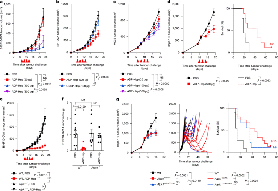
Figure 1. Activation of ALPK1 induces anti-tumor responses in mice
UDSP-Hep: A Potent ALPK1 Agonist That Discriminates Mouse Alpk1 Polymorphisms
To improve the potency of ALPK1 agonists, the team synthesized a series of ADP-Hep analogs. Using NF-κB reporter assays and TIFA phosphorylation analysis, UDP-Hep and CDP-Hep showed approximately 20–40 fold higher activity than ADP-Hep. Further synthesis of the phosphorothioate versions ADSP-Hep and UDSP-Hep yielded even stronger ALPK1 activation. Among them, UDSP-Hep showed an EC₅₀ of 0.0423 μM, about 1/50 that of ADP-Hep (Figure 2a, b).
Its potency in inducing pro-inflammatory factors in human PBMCs and mice was far superior to ADP-Hep (Figure 2d, e). The higher activity of UDSP-Hep is mainly attributed to its greater stability in serum-containing media and mammalian cytosol. Unlike ADP-Hep, UDSP-Hep induces minimal cytokine production in BALB/c and 129 mice and fails to trigger anti-OVA antibody production. This is due to polymorphisms in the Alpk1 gene. ALPK1(C57BL/6) responds well to ADP-Hep, UDP-Hep, and UDSP-Hep, similar to human ALPK1, whereas ALPK1(129) responds only to ADP-Hep (Figure 2f, g). Thus, UDSP-Hep can distinguish ALPK1 variants across mouse strains, suggesting that therapeutic development of ALPK1 agonists should employ C57BL/6 mice.
For C57BL/6 mouse models, our B16F10-OVA cells provide stable antigen presentation and precisely match the requirements of UDSP-Hep related experiments, improving the reliability of research data. Order Now >>
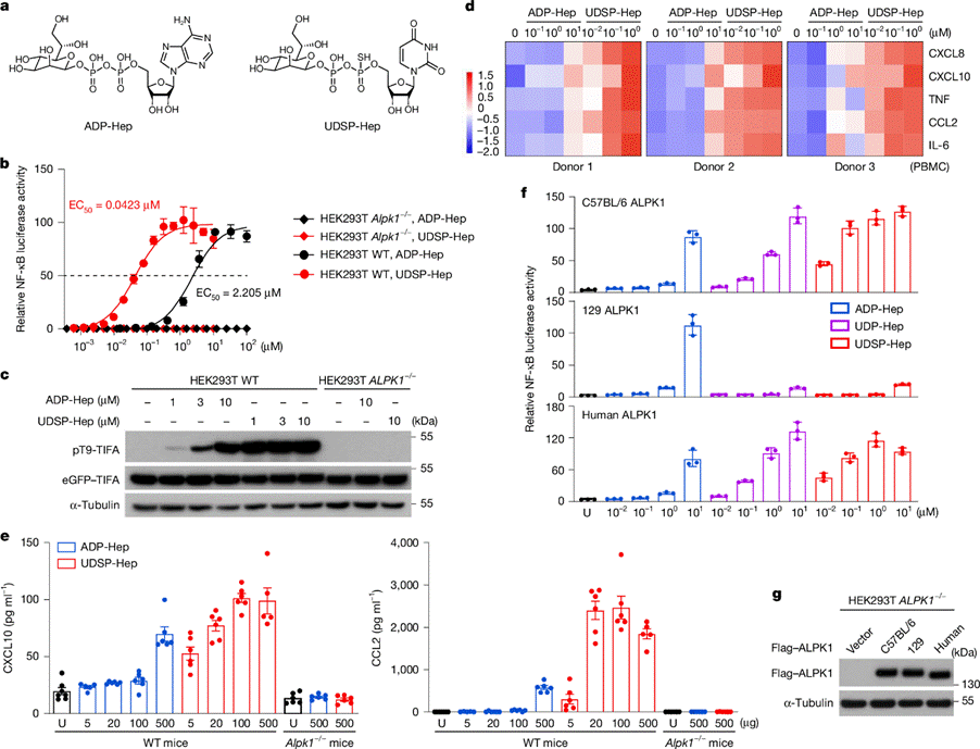
Figure 2. UDSP-Hep is a more potent agonist that discriminates polymorphic Alpk1 alleles in mice
In syngeneic tumor models based on C57BL/6 mice, UDSP-Hep induces near-complete regression of B16F10-OVA, MC38, and Hepa 1-6 tumors at doses much lower than those required for ADP-Hep, and this effect is abolished in Alpk1⁻/⁻ mice. Mice cured of primary tumors by UDSP-Hep are fully protected against tumor re-challenge, indicating the induction of durable anti-tumor immune memory. Bilateral tumor experiments show that injection of UDSP-Hep into one tumor causes equivalent suppression in the contralateral tumor, demonstrating an abscopal effect.
In the MC38 model, combination of UDSP-Hep with anti-CTLA-4 or anti-PD-1 significantly improves tumor control and mouse survival. Even in advanced MC38 tumors, the combination therapy achieves sustained tumor regression in more than half of mice. In naive B16F10 tumors, which are inherently insensitive to single-agent ICI, UDSP-Hep combined with either ICI produces significant tumor control, indicating broad applicability of ALPK1 agonism.
Our 4T1-OVA and B16F10-OVA cells are compatible with multiple mouse tumor models and provide stable and reliable experimental support for both single agonist studies and combination immunotherapy research.
Anti-Tumor Functions of UDSP-Hep: Dependence on CD8⁺ T Cells and Chemokine Regulation
Intratumoral injection of UDSP-Hep rapidly increases intratumoral levels of chemokines including CCL2 and CXCL10 (Figure 3a). Blocking these two factors using neutralizing antibodies significantly reverses the anti-tumor effect of UDSP-Hep, confirming their essential roles in this pathway (Figure 3b). At the tumor microenvironment level, UDSP-Hep treatment leads to marked expansion of CD8⁺ T cells, increased NK cells, and decreased proportion of regulatory T cells, forming a more favorable anti-tumor immune landscape (Figure 3d).
Depletion of CD8⁺ T cells completely abolishes therapeutic efficacy, highlighting the central role of this cell population (Figure 3e). Single-cell transcriptomic analysis further reveals that UDSP-Hep reshapes the immune microenvironment, reduces the expression of T-cell exhaustion-related genes, and enhances activation and survival signals, thereby prolonging anti-tumor immune responses. Mechanistically, UDSP-Hep directly activates dendritic cells in tumor-draining lymph nodes and upregulates the expression of co-stimulatory molecules including CD80 and CD86 (Figure 3h). These activated dendritic cells effectively cross-present antigens, significantly promoting the proliferation and differentiation of antigen-specific CD8⁺ T cells in vitro and in vivo (Figure 4i) and conferring tumor-suppressive activity (Figure 3j). 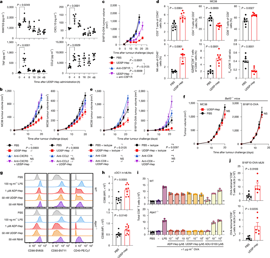
Figure 3. UDSP-Hep induced anti-tumor immunity requires CXCL10 and CCL2 and acts through activation of DCs to drive tumor-specific CD8⁺ T cell expansion
Differences Between UDSP-Hep and STING/TLR Agonists: Unique Anti-Tumor Immune Profiles
Expression profiling shows that ALPK1/TIFA are broadly expressed in cell lines, whereas TLR7/8/9 expression is restricted to hematolymphoid cells, and STING expression is intermediate. Different agonists (UDSP-Hep, R848, ADU-S100) induce distinct cytokine profiles in cells and mice. UDSP-Hep strongly induces CXCL1 and CXCL2, whereas the STING agonist ADU-S100 produces higher levels of IL-6 and TNF. Combination of UDSP-Hep with the STING agonists DMXAA or ADU-S100 results in synergistic anti-tumor effects.
Mechanistically, UDSP-Hep dose-dependently promotes MHC-I presentation of the SIINFEKL epitope on B16F10-OVA cells, whereas ADU-S100 does not. Both agonists stimulate cDC1-mediated cross-priming of CD8⁺ T cells, but UDSP-Hep requires a 10-fold lower concentration. Unlike ADU-S100, which induces T-cell apoptosis, UDSP-Hep and R848 do not cause significant T-cell death. Importantly, after clearing Hepa 1-6 tumors, UDSP-Hep induces a higher proportion of tumor-specific memory T cells, which are key targets of anti-PD-1/PD-L1 blockade.
Summary: Potential of ALPK1-Targeted Immunotherapy and Preferred Experimental Tools
This study confirms that ALPK1 agonism activates CD8⁺ T-cell-mediated anti-tumor immunity and acts synergistically with checkpoint inhibitors. This process depends on macrophages, cDC1, and ALPK1-induced CXCL10/CCL2. Agonists directly activate dendritic cells and promote tumor-specific T-cell expansion.
Compared with STING/TLR pathways, which face challenges in clinical translation, ALPK1 exhibits a broader cellular expression profile and distinct inflammatory characteristics. It not only synergizes with STING/TLR agonists but also stimulates tumor antigen presentation, promotes protective memory T-cell differentiation, and does not induce T-cell apoptosis, demonstrating potential therapeutic advantages. This study lays the foundation for the development of novel ALPK1-targeted immunotherapies.
As preferred tool cells for ALPK1-targeted anti-tumor research, our 4T1-OVA and B16F10-OVA cells meet the needs of diverse experimental scenarios and accelerate the translation of scientific findings.
Reference
Tian, X., Liu, J., Li, Y. et al. Agonists for cytosolic bacterial receptor ALPK1 induce antitumour immunity. Nature 650, 242–250 (2026). https://doi.org/10.1038/s41586-025-09828-9
